

By the time a diabetic foot wound turns black, drains, or refuses to close, the dangerous process may have been building for years.
High blood sugar can quietly damage sensation and blood flow, so a blister you do not feel becomes a wound your body struggles to heal.
That is how a small pressure point can progress to infection, gangrene, and sometimes amputation. The real starting place is usually not the foot. It is the bloodstream.
Before making changes to your diabetes care, medications, exercise routine, or wound treatment, speak with your doctor. A foot problem that looks minor can require prompt professional care when diabetes, poor circulation, or nerve damage is present.
What Denise Means When She Says It Starts in the Bloodstream
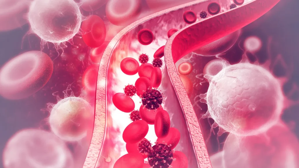
Denise’s point is a teaching shorthand, not a literal rule. Accidents, sudden artery blockages, severe infections, and other conditions can lead to amputations through different routes.
In diabetes, however, the common pathway often begins with prolonged exposure to high blood glucose. Over time, this can damage peripheral nerves and blood vessels. The resulting combination of lost sensation and reduced circulation creates the conditions in which a small injury can become dangerous.
About half of people with diabetes have some form of nerve damage, according to the CDC. Some experience burning, tingling, or numbness, while others have no noticeable symptoms at all.
That absence of pain is not reassuring. It can mean the natural alarm system in your feet is no longer working properly.
The Chain From High Blood Sugar to Amputation
A diabetes related amputation is rarely caused by one isolated event. It usually develops through a chain of overlapping problems.
| Stage | What is happening | What you may notice | Why it matters |
|---|---|---|---|
| Blood vessel injury | High blood sugar and inflammation damage artery walls | Often nothing at first | Plaque can narrow the arteries supplying the legs |
| Nerve injury | Sensory nerves gradually lose function | Tingling, burning, numbness, or no symptoms | A blister, burn, or cut may go unnoticed |
| Repeated pressure | Shoes, calluses, deformities, or walking stress one area | Redness, thick skin, warmth, or a blister | Tissue beneath the skin may begin breaking down |
| Ulcer formation | Skin opens and deeper tissue becomes exposed | A crater, drainage, blood under a callus, or odor | Bacteria can enter the wound |
| Poor healing | Reduced blood flow limits oxygen and immune activity | The wound stays open or enlarges | Infection becomes harder to control |
| Deep infection | Infection reaches deeper tissue or bone | Swelling, redness, drainage, fever, or black tissue | Surgery may be needed to stop spreading infection |
| Amputation | Dead or severely infected tissue cannot be safely preserved | Gangrene or uncontrolled infection | Removing tissue may be necessary to protect the person’s life |
This sequence reflects the pathways described by the CDC, NIDDK, and international diabetic foot guidelines. Peripheral neuropathy, peripheral artery disease, pressure, ulceration, and infection often interact rather than occurring separately.
The First Silent Failure Is Lost Sensation
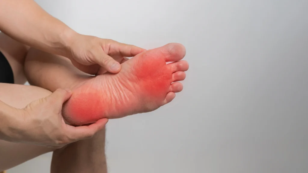
Peripheral neuropathy commonly affects the toes and feet first. It can weaken your ability to detect pain, heat, cold, pressure, and minor trauma.
A person with normal sensation quickly notices a pebble in a shoe. A person with neuropathy may walk on it for hours. The same problem can occur with a tight seam, a curled insole, a new pair of shoes, or a hot floor.
The injury may be small at first. Continued walking repeatedly loads the damaged area, especially beneath the big toe, ball of the foot, heel, or a prominent joint.
Calluses deserve special attention. They are not merely cosmetic when sensation is reduced. Thick callused skin can increase local pressure, and bleeding or tissue breakdown may develop underneath it. International guidelines identify excess callus and early skin lesions as important ulcer risk factors.
Never cut a callus yourself or use an acid based corn remover when you have diabetes or reduced sensation. These products can injure the skin and create an entry point for infection.
The Second Silent Failure Is Poor Circulation
Peripheral artery disease, commonly called PAD, occurs when arteries carrying blood to the legs become narrowed or blocked. Diabetes increases the risk because high blood sugar promotes inflammation and blood vessel damage, making plaque formation more likely.
Reduced circulation means less oxygen reaches injured tissue. It also makes it harder for immune cells and medications to reach an infection effectively.
PAD does not always cause obvious pain. Some people develop calf, thigh, or hip discomfort while walking. Others notice cool feet, color changes, weak pulses, slow growing nails, or wounds that heal poorly.
A painless wound can therefore be especially misleading. Neuropathy hides the injury, while poor circulation prevents it from closing.
Most Amputations Have a Warning Stage
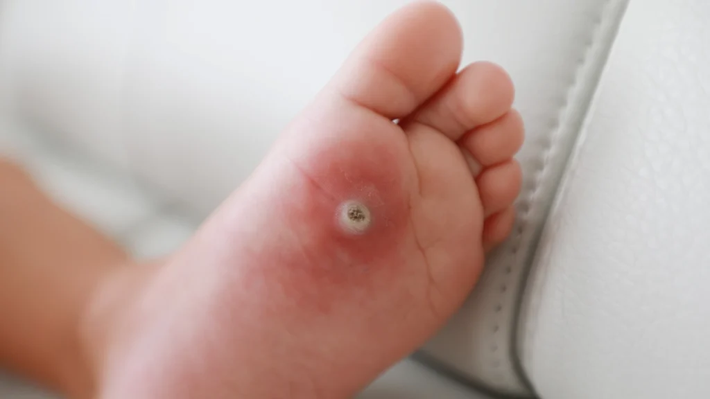
Diabetic foot ulcers precede roughly 80 percent of lower extremity amputations among people with diabetes. That does not mean most ulcers result in amputation.
It means the ulcer is often the critical warning stage at which fast, skilled treatment can change the outcome.
Treatment may involve pressure relief, removal of dead tissue, infection control, specialized dressings, imaging, and an assessment of blood flow. If circulation is severely restricted, vascular specialists may consider a procedure to restore blood flow.
Walking normally on an open plantar ulcer can repeatedly crush healing tissue. This is why pressure relief, often called offloading, is a central part of ulcer treatment rather than an optional extra.
Waiting for pain is one of the most dangerous mistakes. A person with advanced neuropathy may have a deep ulcer or even bone infection with surprisingly little discomfort.
Wound care should be personalized. Do not copy a dressing method, use leftover antibiotics, apply harsh antiseptics, or continue normal activity over an ulcer without professional guidance.
Your A1C Is Important, but It Is Not the Whole Foot Exam
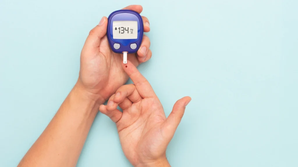
Keeping blood sugar within your agreed target can help prevent nerve damage or slow its progression. It also supports the body’s ability to fight infection and repair tissue.
Still, a glucose reading or A1C result cannot tell you whether you have lost protective sensation, developed PAD, formed a pressure point, or started an ulcer beneath a callus.
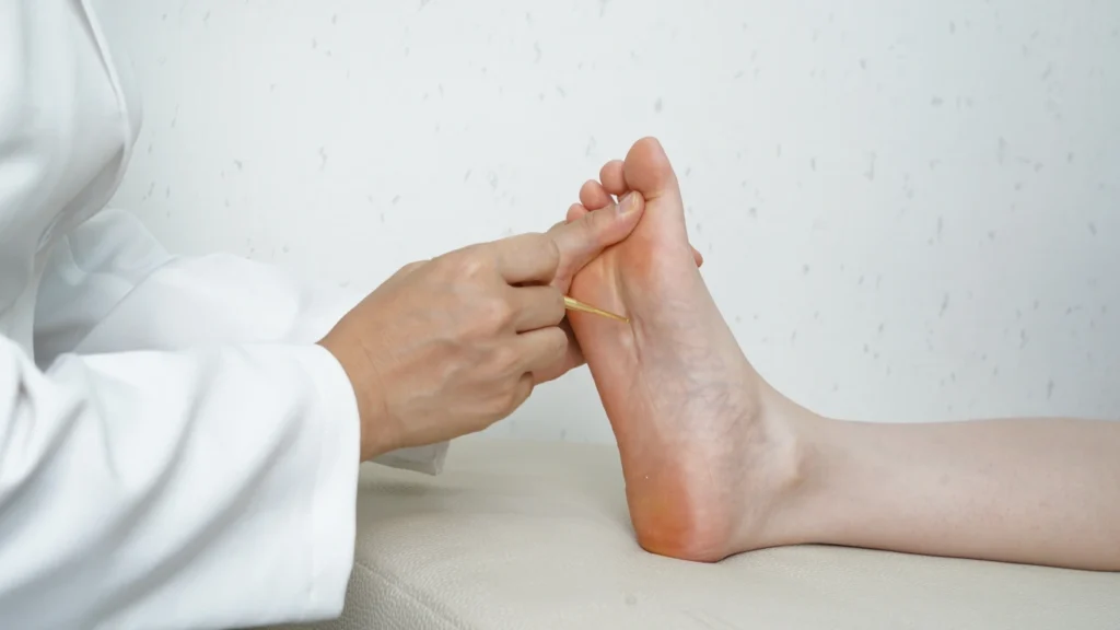
A proper diabetes foot assessment should include skin inspection, foot shape, pulses, circulation symptoms, previous wounds, footwear, and sensation testing. A common clinical tool is a 10 gram monofilament, which checks whether protective sensation is still present.
The International Working Group on the Diabetic Foot recommends screening frequency based on the combination of sensation loss, PAD, deformity, kidney disease, and previous ulcers or amputations.
| Risk level | Typical findings | Suggested professional screening |
|---|---|---|
| Very low | No loss of protective sensation and no PAD | Once a year |
| Low | Loss of protective sensation or PAD | Every 6 to 12 months |
| Moderate | Sensation loss plus PAD, or either condition plus foot deformity | Every 3 to 6 months |
| High | Sensation loss or PAD plus a previous ulcer, previous amputation, or end stage kidney disease | Every 1 to 3 months |
These intervals are professional guideline recommendations, but your clinician may examine you more often based on active symptoms, glucose control, mobility, vision, footwear, or access to home support.
Five Factors That Push Risk Higher
1. A Previous Foot Ulcer
A healed ulcer should be treated as a foot in remission, not a permanent cure. International guidelines report that approximately 40 percent of diabetes related foot ulcers recur within one year after healing.
The old pressure point may still exist. Neuropathy and PAD may also remain, even though the skin has closed.
2. Kidney Disease
Advanced kidney disease is included in the highest IWGDF foot risk category when it occurs with sensation loss or PAD. Kidney disease often travels with severe vascular disease, anemia, swelling, and impaired healing capacity.
3. Smoking
Smoking further reduces blood flow and increases vascular risk. Both the NIDDK and American Heart Association include smoking cessation among the most important steps for protecting circulation.
4. Foot Deformity or Limited Mobility
Bunions, hammertoes, Charcot changes, stiff joints, or an altered walking pattern can shift pressure onto a small area. When sensation is reduced, that pressure may continue without warning pain.
5. Inadequate Footwear
Shoes that are too tight, too loose, worn inside, pointed at the toes, or rough along the lining can cause rubbing and pressure. Guidelines recommend footwear that accommodates the shape of the foot, with specialized shoes or insoles considered for people with deformity or previous plantar ulcers.
The Daily Habit That Can Catch Trouble Early
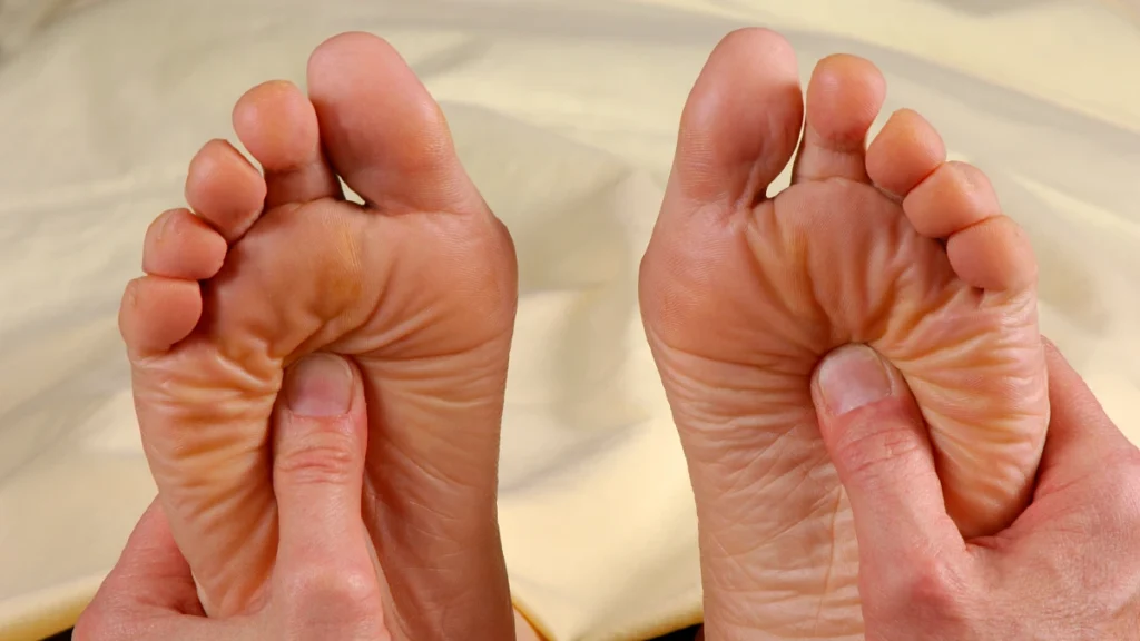
A foot inspection takes about a minute, yet it can reveal a problem before infection or tissue death develops. Check even when your feet feel completely normal, since neuropathy may remove the pain that would otherwise alert you.
Try this every evening
The Daily Habit That Can Catch Trouble Early
A foot inspection takes about a minute, yet it may reveal a problem before infection or tissue death develops. Check even when your feet feel completely normal. Neuropathy may remove the pain that would otherwise warn you.
Do not walk barefoot, even indoors, if you have neuropathy or a high risk foot. The IWGDF specifically advises protective footwear because thin slippers, bare feet, and socks alone do not protect against pressure, heat, or sharp objects.
What to Do When You Find a Problem
| What you see | What to do now | What not to do |
|---|---|---|
| Mild redness after wearing shoes | Remove the shoes, reduce pressure, and recheck the skin soon | Do not keep wearing the same shoes |
| Small blister, cut, or crack | Clean gently, cover with a clean dressing, and contact your care team for advice | Do not pop the blister or apply acids |
| Blood beneath a callus | Arrange prompt podiatry or wound assessment | Do not cut or file deeply into the callus |
| Open ulcer or drainage | Seek professional evaluation promptly | Do not walk normally on it or use leftover antibiotics |
| Black tissue, foul odor, spreading redness, or fever | Seek urgent medical care | Do not wait for pain or a routine appointment |
| Suddenly cold, pale, blue, weak, or numb foot | Seek emergency assessment for possible loss of blood flow | Do not massage it or wait overnight |
The CDC and NIDDK recommend prompt care for wounds that are not healing, color changes, swelling, infection signs, blood beneath callus, and black or foul smelling tissue.
When to Stop Reading and Get Medical Help
Contact a clinician promptly if you notice:
- A blister, cut, or sore that is not beginning to improve within a few days
- New redness, warmth, swelling, drainage, or a bad odor
- Blood beneath a callus or a new warm area on one foot
- A new change in foot color, temperature, shape, or sensation
Seek urgent or emergency care for black tissue, rapidly spreading redness, fever with a foot wound, exposed bone, or a foot that suddenly becomes cold, pale, blue, weak, or numb. These findings may indicate gangrene, deep infection, or severely restricted blood flow.
The Foot Is the Last Link, Not the First
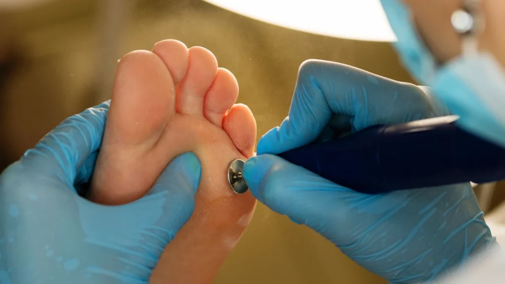
The visible sore may be on the foot, but the process often begins years earlier with injury to nerves and arteries. High blood sugar starts the vulnerability. Neuropathy hides the damage. Pressure opens the skin. PAD delays healing. Infection then turns a local wound into a limb threatening problem.
That chain can be interrupted at several points. Manage blood sugar, blood pressure, cholesterol, smoking, and kidney health with your clinical team. Check your feet daily.
Request a complete professional foot examination at least annually, and more often when sensation loss, PAD, deformity, kidney disease, or a previous ulcer places you at higher risk.
Most importantly, never wait for a diabetic foot wound to hurt before taking it seriously. The absence of pain may be the warning.
This article is for informational purposes only and is not a substitute for professional medical advice, diagnosis, wound assessment, or treatment. Always consult your doctor or a qualified healthcare provider before changing your diet, activity, medications, or foot care routine, especially if you have neuropathy, poor circulation, kidney disease, an active wound, or a previous amputation.